| Current Emergency Medicine, ISSN 0000-0000 online, Open Access |
| Article copyright, the authors; Journal compilation copyright, Curr Emerg Med and Elmer Press Inc |
| Journal website https://cem.elmerpub.com |
Case Report
Volume 1, 2026, e4

An Atypical Presentation of Acute Central Cord Syndrome
Hakan Aka, d  , Sedanur Ozgulb , Turan Cayirc
aDepartment of Neurosurgery, Faculty of Medicine, Kirsehir Ahi Evran University, Kirsehir, Turkey
bDepartment of Emergency Medicine, Kirsehir Training and Research Hospital, Kirsehir, Turkey
cDepartment of Emergency Medicine, Mus Training and Research Hospital, Mus, Turkey
dCorresponding Author: Hakan Ak, Department of Neurosurgery, Faculty of Medicine, Kirsehir Ahi Evran University, Kirsehir, Turkey
Manuscript submitted July 21, 2026, accepted September 15, 2026, published online October 10, 2026
Short title: Atypical Central Cord Syndrome
doi: https://doi.org/10.14740/cem4
| Abstract | ▴Top |
Central cord syndrome (CCS) is the most common form of incomplete spinal cord injury and is classically characterized by greater impairment in the upper extremities compared to the lower extremities. However, atypical clinical presentations may create diagnostic challenges. A 65-year-old male patient presented with cervical trauma following a fall from a height of approximately 2 m. Neurological examination revealed complete motor loss (0/5) in the lower extremities, while upper extremity motor strength was assessed as 3/5 bilaterally. Cervical computed tomography demonstrated a C5 fracture and traumatic disc herniation. Magnetic resonance imaging revealed central cord edema at the C4–C6 levels. No findings of complete cord transection were observed. Based on clinical and radiological findings, the patient was diagnosed with atypical CCS. This case demonstrates that CCS may rarely present with paraplegia, while upper extremity function can be partially preserved. Awareness of such atypical presentations is important for preventing misdiagnosis and determining appropriate treatment strategies.
Keywords: Central cord syndrome; Cervical trauma; Paraplegia; Incomplete spinal cord injury
| Introduction | ▴Top |
Central cord syndrome (CCS) is the most common form of incomplete spinal cord injury and most frequently results from a cervical hyperextension mechanism. Owing to the high prevalence of cervical spondylotic stenosis, CCS is more commonly observed in older adults, as it may develop even after low-energy falls or minor hyperextension injuries [1]. Clinically, it is characterized by disproportionately greater motor weakness in the upper extremities than in the lower extremities. Although this neurological pattern was traditionally attributed to the somatotopic organization of the corticospinal tracts within the spinal cord, recent studies have questioned this traditional view and provided evidence challenging the concept of a strict somatotopic organization [2, 3].
Pathophysiologically, CCS is associated with edema, contusion, or hemorrhage in the central portion of the spinal cord following cervical hyperextension trauma. The pathophysiology of CCS is complex and remains incompletely understood. Although the characteristic neurological pattern was traditionally attributed to a somatotopic organization of the corticospinal tract, this anatomical model has been challenged by recent neuroanatomical and clinical evidence [2, 3]. Current evidence suggests that the clinical manifestations of CCS cannot be adequately explained by a strict medial-to-lateral organization of corticospinal tract fibers and may instead reflect the location and extent of spinal cord injury, including the effects of mechanical compression, edema, and axonal injury [2, 3]. Therefore, upper extremity weakness predominates in classical presentation.
However, recent studies have shown that the clinical spectrum of CCS extends beyond the classical definition and may present with different neurological patterns. In rare cases, atypical presentations with predominant lower extremity involvement or paraplegia may occur. Such cases can lead to diagnostic confusion in patients presenting with paraplegia after cervical trauma and may be misinterpreted as complete spinal cord injury or lower-level lesions [3].
Magnetic resonance imaging (MRI) is the most valuable modality in the diagnosis of CCS and plays a critical role in demonstrating central spinal cord edema or structural lesions. Early diagnosis and appropriate treatment are crucial for neurological recovery [4].
In this report, we present a rare case of atypical CCS in a patient who presented with paraplegia following cervical trauma, with partially preserved upper extremity motor function. The aim is to highlight atypical clinical presentations of CCS and emphasize their consideration in the differential diagnosis.
| Case Report | ▴Top |
A 65-year-old man presented to the emergency department after falling from a height of approximately 2 m. He had no known comorbidities. On admission, he was conscious (Glasgow Coma Scale score: 15) and hemodynamically stable. Neurological examination revealed complete motor paralysis of both lower extremities (Medical Research Council (MRC) grade 0/5) and bilateral upper extremity weakness with an MRC grade of 3/5. Sensory examination demonstrated diminished pain and temperature sensation below the C5 dermatome, while light touch sensation was preserved. Deep tendon reflexes were hypoactive. Voluntary anal contraction was preserved, indicating sacral motor sparing and supporting the diagnosis of a motor-incomplete spinal cord injury.
Cervical computed tomography (CT) demonstrated an anterior endplate fracture of the C5 vertebral body. In addition, findings suggestive of an acute traumatic C5–6 intervertebral disc herniation were observed (Fig. 1a, b). No significant vertebral dislocation or facet joint dislocation was identified. Cervical MRI revealed centrally located intramedullary T2-weighted hyperintense signal changes consistent with spinal cord edema extending from C4 to C6, as well as an acute C5–6 disc herniation (Fig. 2a, b).
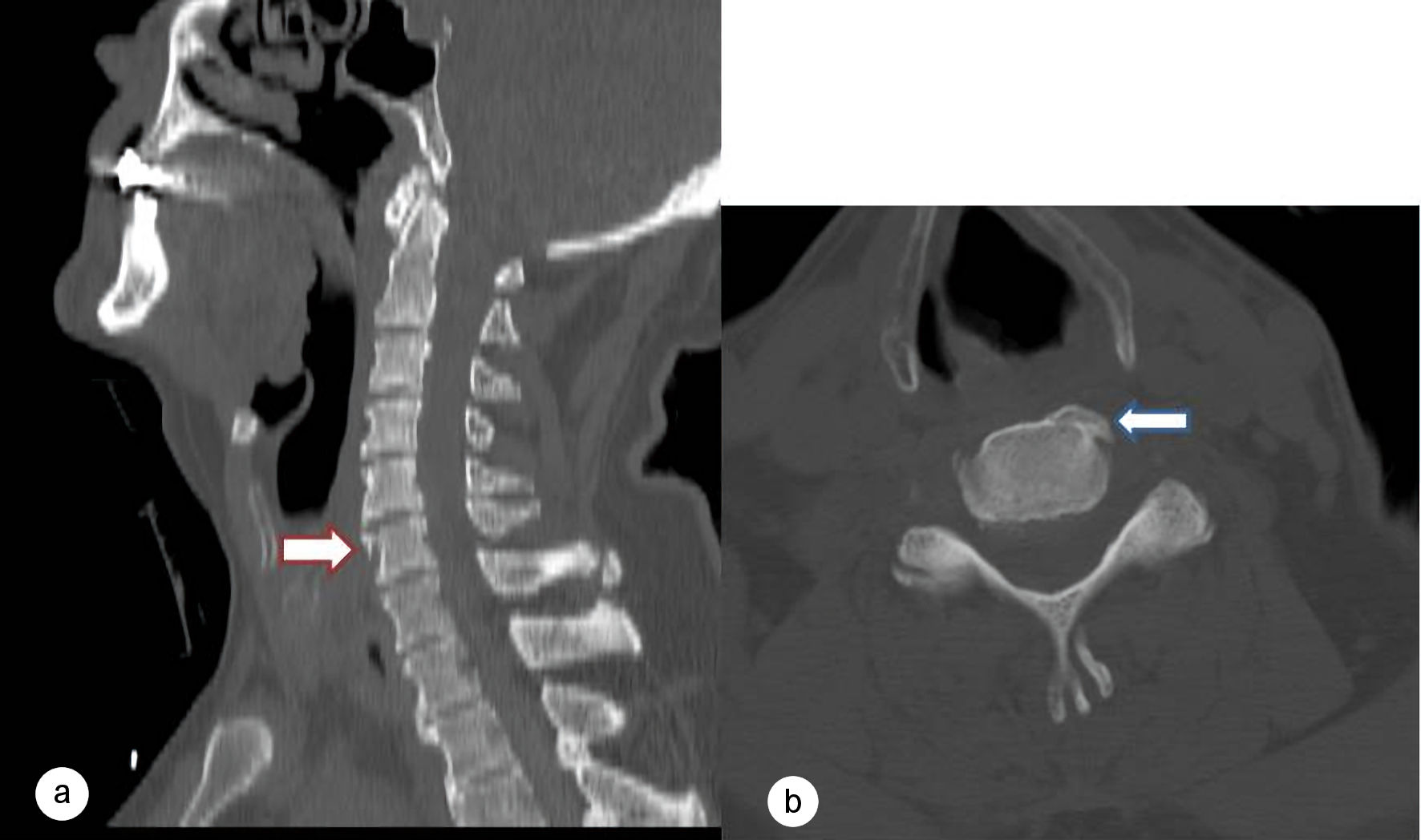 Click for large image | Figure 1. Preoperative cervical computed tomography (CT). (a) Sagittal CT image demonstrating an anterior endplate fracture of the C5 vertebral body (red arrow). (b) Axial CT image showing the C5 vertebral body fracture without evidence of posterior element or facet joint injury (white arrow). |
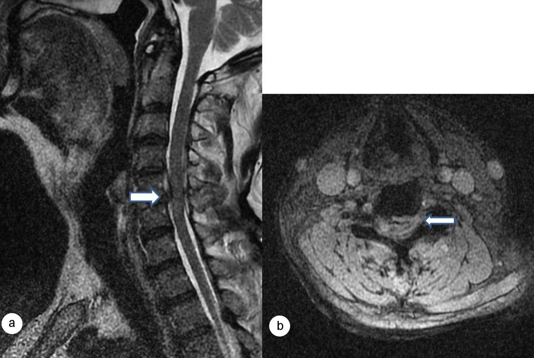 Click for large image | Figure 2. Preoperative cervical magnetic resonance imaging (MRI). (a) Sagittal T2-weighted image demonstrating an acute C5–6 traumatic disc herniation causing ventral spinal cord compression with intramedullary T2 hyperintensity consistent with central cord edema (white arrow). (b) Axial T2-weighted image demonstrating centrally located spinal cord edema at the C5 level (white arrow). |
Given the presence of an acute traumatic C5–6 disc herniation causing significant compression of the spinal cord, together with the severe neurological deficit characterized by complete motor paralysis of both lower extremities and bilateral upper extremity weakness, surgical decompression was considered necessary. The patient underwent C5–6 anterior microsurgical discectomy followed by anterior cervical stabilization (Fig. 3).
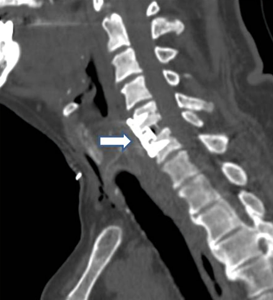 Click for large image | Figure 3. Postoperative sagittal cervical computed tomography (CT) demonstrating successful anterior cervical discectomy and fusion (ACDF) at the C5–6 level with appropriate placement of the interbody cage and anterior cervical plate (white arrow). |
In the immediate postoperative period, neurological examination demonstrated preserved bilateral upper extremity motor strength (MRC grade 3/5), while lower extremity motor strength improved to MRC grade 1/5. At the first postoperative week, upper extremity motor strength had improved to MRC grade 4/5, and lower extremity motor strength had improved to MRC grade 2/5.
Postoperatively, the patient remained clinically stable, although he was unable to achieve independent mobilization because of persistent lower extremity weakness. The early rehabilitation program was continued during the postoperative period. At the first postoperative week, upper extremity motor strength had improved to MRC grade 4/5, while lower extremity motor strength had improved to MRC grade 2/5. Enoxaparin 0.6 mL (6,000 anti-Xa IU/60 mg) twice daily was initiated 24 h after surgery for thromboprophylaxis. Approximately 3 weeks after surgery, the patient developed a decrease in oxygen saturation on room air. In view of the new-onset oxygen desaturation, pulmonary CT angiography was performed, which confirmed pulmonary embolism. Despite appropriate management, the patient subsequently died due to pulmonary embolism.
| Discussion | ▴Top |
CCS is the most common incomplete spinal cord syndrome, whereas anterior cord syndrome, Brown-Sequard syndrome, posterior cord syndrome, conus medullaris syndrome, and cauda equina syndrome are encountered less frequently. CCS is the most common form of incomplete spinal cord injury; however, its reported frequency varies substantially depending on the diagnostic criteria and study population [5, 6]. However, no data are currently available in the literature regarding the actual or approximate incidence of atypical CCS or its variants.
The most common mechanism of CCS is cervical hyperextension injury, particularly in elderly patients with underlying cervical spondylosis [1, 7]. In this setting, compression of the spinal cord between anterior osteophytes and the posterior ligamentous structures may result in central cord edema and/or intramedullary hemorrhage [3, 4]. In contrast, the present case involved a C5 vertebral fracture associated with an acute traumatic C5–6 disc herniation following a fall from height. The acute disc herniation likely produced direct mechanical compression of the spinal cord, while the accompanying spinal cord edema may have exacerbated secondary neural injury, thereby contributing to the patient’s atypical neurological presentation.
Although CCS classically presents with greater motor impairment in the upper extremities than in the lower extremities, considerable variability, including atypical clinical presentations, has been reported [6, 8]. Unilateral motor weakness has also been described as an atypical presentation [8]. However, the occurrence and pathophysiological basis of presentations with predominant lower-extremity involvement or paraplegia remain poorly defined. The underlying pathophysiological mechanisms remain incompletely understood but are thought to involve variable degrees of spinal cord edema, vascular compromise, and selective involvement of corticospinal tract fibers by associated structural lesions [3].
In addition to CCS, other incomplete spinal cord syndromes include anterior cord syndrome, posterior cord syndrome, Brown-Sequard syndrome, conus medullaris syndrome, and cauda equina syndrome. Considering the level and characteristics of the injury in our patient, anterior cord syndrome represents the most important differential diagnosis that should be carefully considered. In anterior cord syndrome, acute neurological manifestations may include bilateral flaccid paraparesis, quadriparesis, or paralysis, accompanied by loss of pain and temperature sensation, while proprioception, vibration, fine touch, and two-point discrimination are relatively preserved. Among the acute clinical manifestations of anterior cord syndrome, autonomic dysfunction accompanied by hypotension, bradycardia, impaired thermoregulation, respiratory failure, and spinal shock may also occur [9]. In our patient, vital signs remained stable throughout the clinical course. Neurological examination revealed predominantly lower-extremity motor impairment, with partial preservation of motor strength in the upper extremities. Sensory examination demonstrated preservation of superficial sensation.
Brown-Sequard syndrome represents another important differential diagnosis and results from injury involving one half of the spinal cord (hemicord). It is classically characterized by ipsilateral impairment of motor function, vibration, and proprioception, accompanied by contralateral loss of pain and temperature sensation [10]. The neurological findings observed in our patient were not consistent with the characteristic clinical pattern of Brown-Sequard syndrome.
Following comprehensive sensory and motor neurological examination and determination of the neurological level, patients with spinal cord injury are classified into five categories according to the ASIA Impairment Scale (AIS), ranging from AIS A to AIS E. Patients with no preserved sensory or motor function in the sacral segments (S4–S5) are classified as AIS A, indicating a complete spinal cord injury. Preservation of sensory function at S4–S5, including light touch, pin-prick, or deep anal pressure, in the absence of preserved motor function more than three neurological levels below the motor level is consistent with a sensory-incomplete injury (AIS B). A motor-incomplete injury is defined by the presence of voluntary motor function in the most caudal sacral segments or by fulfillment of the criteria for sensory incompleteness together with preservation of motor function more than three levels below the ipsilateral motor level. Non-key muscles may also contribute to the determination of motor completeness. Patients with preserved muscle strength of ≥ 3 in fewer than half of the key muscles below the neurological level are classified as AIS C, whereas those with preserved strength of ≥ 3 in at least half of the key muscles below the neurological level are classified as AIS D. Patients with a previous neurological deficit who subsequently demonstrate normal sensory and motor function according to the International Standards for Neurological Classification of Spinal Cord Injury (ISNCSCI) are classified as AIS E [11].
According to the ISNCSCI, the patient demonstrated preserved sacral motor function, as evidenced by voluntary anal contraction, indicating a motor-incomplete spinal cord injury. Motor function was also preserved below the neurological level, including T1 motor function (MRC grade 3/5). However, fewer than half of the key muscles below the neurological level demonstrated muscle strength of ≥ 3/5, while the remaining key muscles had muscle strength ranging from 0 to 2/5. Based on these neurological findings, the patient was classified as AIS C.
MRI plays a pivotal role in the diagnosis of spinal cord injury by accurately demonstrating intramedullary edema, hemorrhage, and associated soft tissue injuries [12, 13]. In the present case, centrally located spinal cord edema extending from C4 to C6 correlated well with the patient’s neurological deficits and supported the diagnosis. CT, however, remains indispensable for identifying associated osseous injuries, particularly fractures involving the vertebral body and posterior elements [12, 13]. In our patient, CT demonstrated an anterior endplate fracture of the C5 vertebral body without evidence of posterior element injury.
From a clinical standpoint, CCS generally carries a more favorable neurological prognosis than complete spinal cord injury, particularly in patients with preserved motor function. Early diagnosis, appropriate cervical immobilization, timely surgical decompression when indicated, and comprehensive rehabilitation are key factors associated with improved functional recovery [1, 4]. In the present case, neurological improvement was observed during the early postoperative period. However, despite this favorable neurological course, the patient died 3 weeks after surgery due to pulmonary embolism, a systemic complication unrelated to the neurological recovery.
Conclusions
In conclusion, this case highlights that atypical presentations of CCS should be considered in the differential diagnosis of patients presenting with paraplegia following cervical trauma. Awareness of these uncommon clinical patterns is essential to avoid misdiagnosis, facilitate timely use of appropriate imaging modalities, and guide appropriate management based on the individual clinical and radiological findings.
Acknowledgments
None to declare.
Financial Disclosure
The authors received no financial support or funding for the research, authorship, and/or publication of this article.
Conflict of Interest
The authors declare that they have no conflict of interest.
Informed Consent
Informed consent was obtained from the patient’s legal representative for publication of this case report and accompanying images.
Author Contributions
HA conceived and designed the study. HA and SO collected the clinical data. SO performed the literature review and drafted the manuscript. TC critically revised the manuscript for important intellectual content. All authors read and approved the final version of the manuscript.
Data Availability
The data supporting the findings of this study are available from the corresponding author upon reasonable request.
| References | ▴Top |
- Ameer MA, Tessler J, Munakomi S, Gillis C. Central cord syndrome. In: StatPearls [Internet]. Treasure Island (FL): StatPearls Publishing; 2023.
- Levi AD, Schwab JM. A critical reappraisal of corticospinal tract somatotopy and its role in traumatic cervical spinal cord syndromes. J Neurosurg Spine. 2022;36(4):653-659.
doi pubmed - Shakil H, Santaguida C, Wilson JR, Farhadi HF, Levi AD, Wilcox JT. Pathophysiology and surgical decision-making in central cord syndrome and degenerative cervical myelopathy: correcting the somatotopic fallacy. Front Neurol. 2023;14:1276399.
doi pubmed - Avila MJ, Hurlbert RJ. Central cord syndrome redefined. Neurosurg Clin N Am. 2021;32(3):353-363.
doi pubmed - Bosch A, Stauffer ES, Nickel VL. Incomplete traumatic quadriplegia. A ten-year review. JAMA. 1971;216(3):473-478.
pubmed - Engel-Haber E, Snider B, Botticello A, Eren F, Kirshblum S. Clinical subsets of central cord syndrome: Is it a distinct entity from other forms of incomplete tetraplegia for research? J Neurotrauma. 2024;41(17-18):2133-2145.
doi pubmed - Martinez-Palacios K, Rubiano AM, Demetriades AK, Vasquez-Garcia S. Traumatic central cord Syndrome: An integrated neurosurgical and neurocritical care perspective. Brain Spine. 2025;5:104281.
doi pubmed - Shaw EV. Central cord syndrome presenting as unilateral weakness. Am J Emerg Med. 1995;13(1):41-42.
doi pubmed - Sandoval JI, De Jesus O. Anterior spinal artery syndrome. In: StatPearls. Treasure Island (FL) ineligible companies. 2024.
pubmed - Shams S, Farrell CL, Arain A. Brown-Sequard syndrome. In: StatPearls. Treasure Island (FL) ineligible companies. 2024.
pubmed - http://asia-spinalinjury.org/wp-content/uploads/2016/02/ International_Stds_Diagram_Worksheet.pdf.
- Kumar Y, Hayashi D. Role of magnetic resonance imaging in acute spinal trauma: a pictorial review. BMC Musculoskelet Disord. 2016;17:310.
doi pubmed - Hussain O, Kaushal M, Agarwal N, Kurpad S, Shabani S. The role of magnetic resonance imaging and computed tomography in spinal cord injury. Life (Basel). 2023;13(8):1680.
doi pubmed
This article is distributed under the terms of the Creative Commons Attribution 4.0 International License (CC BY 4.0), which permits unrestricted use, distribution, and reproduction in any medium, including commercial use, provided the original work is properly cited.
Current Emergency Medicine is published by Elmer Press Inc.