Current Emergency Medicine, ISSN NoIssnPrint print, 0000-0000 online, Open Access
Article copyright, the authors; Journal compilation copyright, Curr Emerg Med and Elmer Press Inc
Journal website https://cem.elmerpub.com

Case Report

Volume 1, 2026, e4


An Atypical Presentation of Acute Central Cord Syndrome

Figures

↓  Figure 1. Preoperative cervical computed tomography (CT). (a) Sagittal CT image demonstrating an anterior endplate fracture of the C5 vertebral body (red arrow). (b) Axial CT image showing the C5 vertebral body fracture without evidence of posterior element or facet joint injury (white arrow).
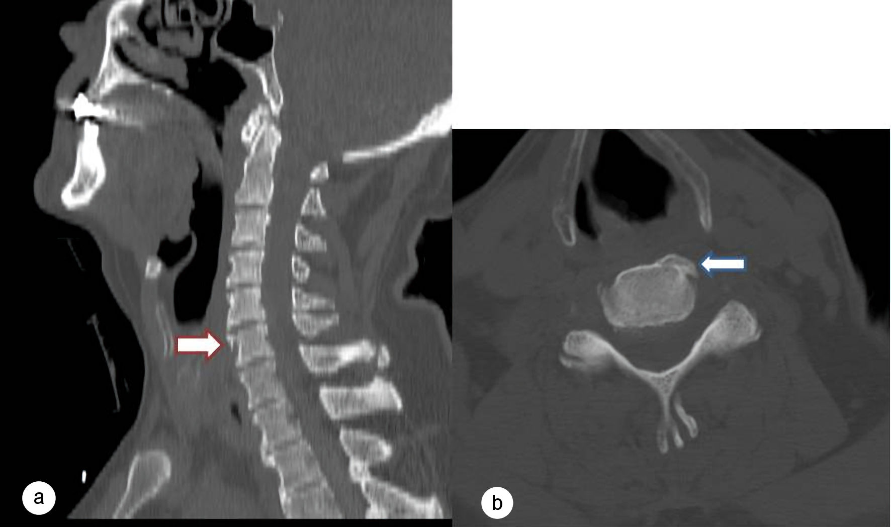
Figure 1.
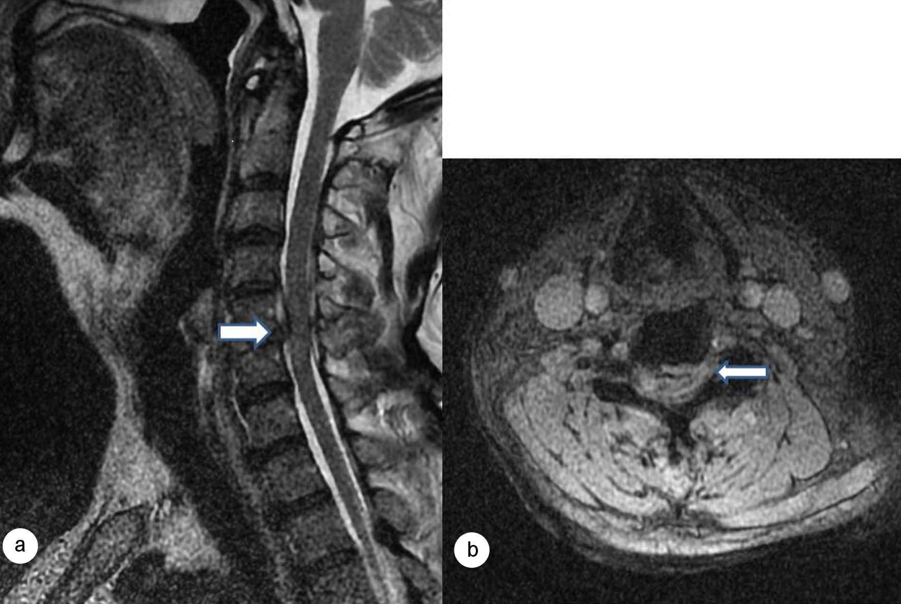
↓  Figure 2. Preoperative cervical magnetic resonance imaging (MRI). (a) Sagittal T2-weighted image demonstrating an acute C5–6 traumatic disc herniation causing ventral spinal cord compression with intramedullary T2 hyperintensity consistent with central cord edema (white arrow). (b) Axial T2-weighted image demonstrating centrally located spinal cord edema at the C5 level (white arrow).
Figure 2.
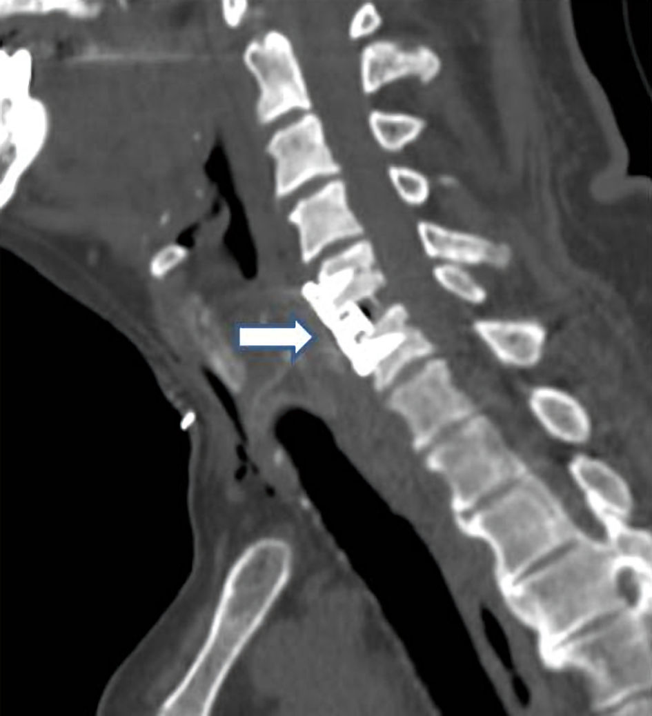
↓  Figure 3. Postoperative sagittal cervical computed tomography (CT) demonstrating successful anterior cervical discectomy and fusion (ACDF) at the C5–6 level with appropriate placement of the interbody cage and anterior cervical plate (white arrow).
Figure 3.